 |
 |
|
|
|
Single-particle analysis for proliferation index detection |
|
|
|
The aim is to identify, isolate and analyze a micrometric particle with an isotope that stands out among thousands of others.
In nuclear proliferation inspections, several applications take priority, including:
|
 |
detection of clandestine activity, |
|
|
identification of traffic in fissile materials, |
|
|
expert assessments to support international institutions (IAEA, Euratom). |
|
|
|
|
 Sample treatment in clean rooms Sample treatment in clean rooms |
|
|
|
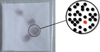
Unlike full chemical treatment of a given sample, which provides an average value for its overall isotopic composition, single-particle analysis provides the isotopic composition of each single particle.

 |
Samples are treated in clean rooms to prevent any pollution or cross-contamination. |
|
|
|
The red dot represents a particle containing fissile atoms 235U
and/or 239Pu |
|
|
|
|
|
Particle irradiation and detection |
|
|
|
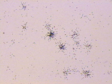
The "stars" formed by the impact of fission fragments during neutron irradiation of particles in the reactor enable identification of the sought particles, which are those containing fissile atoms.
 |
|
 |
|
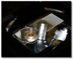
Neutron irradiation in a reactor. |
|
|
|
Fission trace observation through optical microscope. |
|
| |
|
|
 |
|
|
|
Neutron irradiation shuttle. |
|
|
|
Each "star" enables the location of particles containing fissile atoms. |
|
|
|
|
|
Detected particle analysis |
|
|
|
The Military Applications Division (DAM) teams have three analysis methods at their disposal that can be combined. |
|
|
|
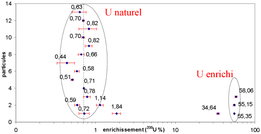
Micro-handling of isotopic analysis by TIMS |
|
(Thermal Ionization Mass Spectrometry).
|
Example of method application: detection of two families with different enrichment: natural uranium 235U/238U = 0,72, highly enriched uranium 235U/238U = 55. |
|
|
|
|
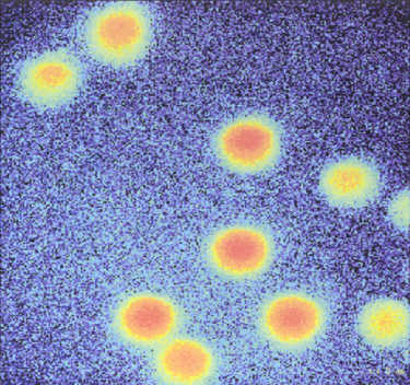
Ionic imaging by SIMS (Secondary
Ion Mass Spectrometry). |
|
|
|
SIMS provides the capability for resolving fine-scale values for isotope analysis without needing to irradiate the sample prior to analysis in some cases. SIMS also provides an ionic picture.
|
238U signals of 1µm diameter UO2 particles. |
|
|
|
|
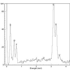
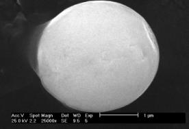
Morphology and particle analysis using an X analyzer coupled with a SEM (Scanning Electron Microscopy). |
|
|
|
Particles containing uranium can be detected semi-automatically by electron microscopy. For each particle detected in this way, the analysis of X-rays emitted on electron bombardment in SEM enables identification of the particle's chemical components. This information can sometimes help to determine the origin of the particle.
 |
|
 |
|
Typical UO2F2 particle, released through certain uranium enrichment operations, identified by X-ray. |
|
|
|
Morphological analysis |
|
|
| |
|